
This year, the American Association for Cancer Research (AACR) Annual Meeting 2021 is taking place over two weeks, starting on 10-15 April 2021 and continuing on 17-21 May 2021. The AACR Annual Meeting is the premier event for cancer researchers, and gathers top professionals from around the world to share the state of the art in the fight against cancer.
The IMMUcan consortium partners working on Broad Profiling (Work Package 4.1) have prepared a poster for the AACR 2021, which will be exhibited online from 10 April 2021. The poster – A toolbox enabling a data-driven selection of regions of interest in tumor tissue sections for imaging mass cytometry analysis – was developed together with partners from EORTC, IBBL, SIB Swiss, the University of Zurich and CHUV.
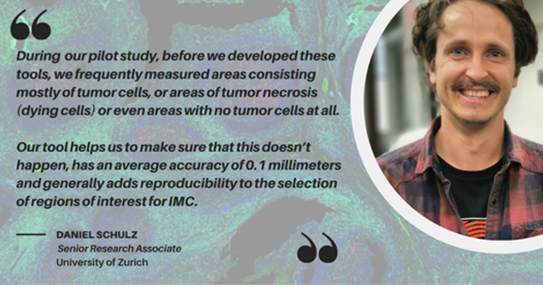
To better understand the significance of the research presented by the team, we sat down for an interview with Daniel Schulz, Senior Research Associate at the University of Zurich, and the lead author of the poster.
Thank you Daniel for speaking with me today. Can you tell us a little bit about your work and the poster?
The IMMUcan project Broad Profiling team generates multiplexed immunofluorescence (mIF), imaging mass cytometry (IMC) and whole exome sequencing/ RNA sequencing data for thousands of cancer patients.
Intriguingly, mIF generates images of whole tumor sections and IMC generates highly multiplexed images for specific regions of each tumor. One of the problems we stumbled across was the selection of these so-called “Regions of interest” for IMC.
So how did you end up selecting those regions of interest?
To answer this question, we have to take a few steps back. Essentially, when we begin, we are more or less ‘blind’ about where to measure.
What we have done is to use the information from our colleagues in Lausanne (CHUV and Swiss Institute of Bioinformatics), which provide mIF data, to decide where to measure, which is possible because we study adjacent tumor sections.
One of the mIF panels that our colleagues from CHUV use contains information about major immune cells (like B cells, T cells, macrophages, neutrophils and tumor cells). We then used this data as the basis for the selection of regions of interest. This is the basis for the poster that we are presenting at this year’s AACR.

What does the tool help you do?
We built a webtool to display some pre-calculated summary statistics like tumor content and immune cell content for all possible regions of the tumor. The tool allows us to ‘zoom in’ on the whole tumor image and identify regions of interest based on the image and the statistics.
We can then select regions, usually 3-4, that contain a certain set of immune cells, and which capture the immune cell heterogeneity present in individual tumors.
Once we selected regions of interest we can download the data. We can then use this data to retrieve the coordinates for measurements with IMC.
And what does this help you to achieve?
There are two main benefits to the tool we developed. When we measure a tumor sample, we measure about 2 x 40 different markers per tumor section. However, we are very focused on the immune system and our panels only contain a handful of markers that target tumor cells.
During our pilot study, before we developed these tools, we frequently measured areas consisting mostly of tumor cells, or areas of tumor necrosis (dying cells) or even areas with no tumor cells at all. Our tool helps us to make sure that this doesn’t happen. It has an average accuracy of 0.1 millimetres and generally adds reproducibility to the selection of regions of interest for IMC.
We can also zoom in and focus on specific regions. One could even envision that in the future researchers could mark their regions of interest using the web tool, send us the ‘coordinates,’ and we would then run it through our pipeline and measure the region of interest for them.
Another benefit is that once we have associated the two modalities (the general tumor image and the ‘zoomed in’ version), we can link the data for analyses.
Has this been done by other researchers?
There are several ways to achieve what we’ve done. But what is special about the way that we have done it for the IMMUcan consortium, is that the data comes from different technologies in different places and from different partners.
Here, we have standardised this process from a partner and the information is now available on the web tool – its independent of where you are, it is hosted on a web server and you can still obtain all the information that you need.
Also, an alignment tool that can handle this kind and this amount of data wasn’t available yet, so we also built a plug-in for napari to help us process the whole tumor mIF images, which can be computationally very intense.
Now bringing it back to the crux of the project – helping the patients: how does this ultimately impact patients, or a person that donates their sample?
Ultimately, we hope that this type of information can have the potential to help us personalise treatment to someone’s specific tumor make-up. And some of the data is actually returned to the physicians as timely molecular reports (on average, within 6 weeks of patient enrolment).
However, there is still so much that we don’t know about cancer and especially the importance of the immune system, which is why studies like these are needed to deepen our understanding of the underlying tumor biology.
Especially with the strength of all approaches – we obtain a lot of information. From mIF about the whole tumor (is it overall inflamed? Does it have good or bad phenotypes); from RNA-Seq and whole exome sequencing we can derive molecular and genetic profiles; the ‘zoomed in’ information from IMC can help us to deeply characterise specific regions of tumors, and have a more complete picture.
In the end, together with our bioinformatics partners we will integrate all these datasets and associate them with clinical features from our patients (e.g. treatment success) to identify the most predictive parameters, which can then be further investigated.
The last day to access the conference materials and posters will be 21 June 2021. You can view the IMMUcan submission at this hyperlink.
IMMUcan aims to study the tumour microenvironment in a bid to gain a deeper understanding of how the immune system and cancer cells interact at the molecular level. The project will also aim to develop a sustainable data platform and legal framework where partners can pursue their own independent investigations.
The IMMUcan project has received funding from the Innovative Medicines Initiative 2 Joint Undertaking under grant agreement No 821558. This Joint Undertaking receives support from the European Union’s Horizon 2020 research and innovation programme and EFPIA.